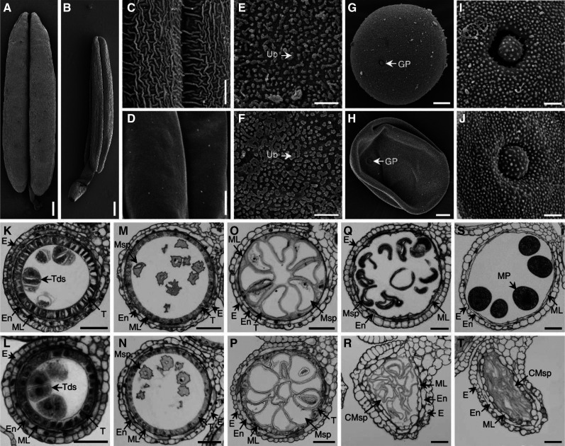
Figure 2.
SEM analysis and transverse section of wild-type and ipe2 anthers. A and B, Mature anthers of wild type (A) and ipe2 (B). C and D, Mature anther outer surfaces of wild type (C) and ipe2 (D). E and F, Mature anther inner surfaces of wild type (E) and ipe2 (F). G and H, Mature pollen grains of wild type (G) and ipe2 (H). I and J, Mature pollen grains outer surfaces with germination apertures of wild type (I) and ipe2 (J). K and L, Anthers of wild type (K) and ipe2 (L) at tetrad stage. M and N, Anthers of wild type (M) and ipe2 (N) at microspore release stage. O and P, Anthers of wild type (O) and ipe2 (P) at large vacuole stage. Q and R, Anthers of wild type (Q) and ipe2 (R) at binucleate stage. S and T, Anthers of wild type (S) and ipe2 (T) at mature pollen stage. CMSp, cCollapsed microspore; E, epidermis; En, endothecium; GP, germination pore; ML, middle layer; MP, mature pollen; MSp, microspore; T, tapetum; Tds, tetrads; Ub, Ubisch body. Scale bars= 400 μm in (A and B), 6 μm (C and D), 4 μm (E and F), 10 μm (G and H), 2 μm (I and J), and 50 μm (K–T).

