Figure 6.
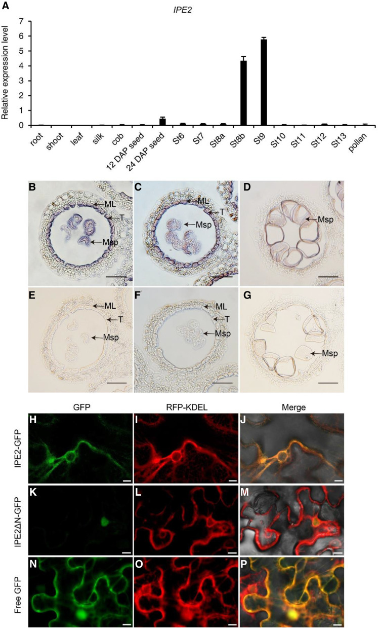
Spatiotemporal expression pattern and subcellular localization of IPE2. A, Expression pattern of IPE2 by RT-qPCR. DAP, Days after pollination. Error bars indicate sd (n = 3). B to G, RNA in situ hybridization of the wild-type anthers using an IPE2-specific antisense (B–D) and a negative control sense probe (E–G). Hybridization signal at stage 9, early uninucleate microspore stage, with winkled microspores (B) and rounded microspores (C). Hybridization signal at stage 10 (D). ML, Middle layer; Msp, microspore; T, tapetum. Scale bars = 50 μm. H to P, Subcellular localization of IPE2 with tobacco transformation showing IPE2-GFP and RFP-KDEL colocalization (H–J), nuclear localization of IPE2ΔN-GFP (K–M), and the control empty construct with GFP (N–P). Scale bars = 10 μm.

