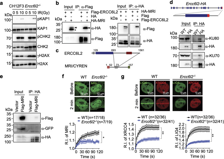
Fig. 4. ERCC6L2 is a component of end-joining machinery.
a Phosphorylation of ATM substrates was examined by western blot. Indicated CH12F3 cell lines were treated with IR at different dosages, and representative blots are showed from three repeats. b Co-immunoprecipitation (Co-IP) of ERCC6L2 and MRI/CYREN in HEK293T cells. The panels show western blots with anti-Flag or anti-HA antibodies of total extract (input) and IP samples. Arrows indicate bands of HA-MRI. Asterisk marks an unknown form of HA-MRI that does not interact with ERCC6L2. c Schematic illustration of ERCC6L2-MRI interaction domains. d Inputs and anti-HA-immunoprecipitated samples from CSR-activated B cells were assayed for NHEJ subunits. WT: B cells from wild-type mice; HA: B cells from Ercc6l2-HA knock-in mice. Knock-in strategy of Ercc6l2-HA mice is showed at top. e Inputs and anti-HA-immunoprecipitated samples from Ercc6l2-HA knock-in CSR-activated B cells with retroviral-expressed Flag-tagged MRI. Flag-MRI-IRES-GFP was ectopically expressed in CSR-activated B cells with an empty retroviral vector (IRES-GFP) as control. IRES internal ribosome entry site. f Recruitment of MRI to DNA damage site in WT and ERCC6L2-deficient MEFs. Upper, total numbers of micro-irradiated cells are listed alone with numbers of cells showed detectable MRI accumulation. Percentage of cells with MRI accumulation are showed in parenthesis. Exampled images of GFP-MRI recruitment are showed at lower. Relative intensity (R.I.) of GFP-MRI at DNA damages are calculated. g Recruitment of XRCC4/LIG4 to DNA damage site in WT and ERCC6L2-deficient MEFs. GFP-LIG4 and mCherry-XRCC4 were co-expressed in cells, and the panel is illustrated as in (f). Data are represented as mean ± SEM in (f) and (g). A t-test was applied as described in the “Materials and Methods” section. **p < 0.01; *p < 0.05.

