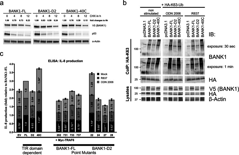
Fig. 4.
Functional analyses of BANK1 proteins. a Protein stability of BANK1–FL, BANK1–D2, and BANK1–40C was studied in nonstimulated U2OS cells by treating cells with 100 µg/ml cycloheximide (CHX) for a time frame of 12 h. β-Actin was used as a housekeeping protein to normalize samples by ImageJ. See also Supplementary Fig. S10. b K63-linked polyubiquitination of BANK1–FL and -D2 and BANK1–40C was tested in nonstimulated HepG2 cells and in cells stimulated O/N with 2 µM ODN 2006 and 8 µg/ml R837. Pull-downs were performed with an anti-HA antibody O/N at 4 °C, and eluates of immunoprecipitates were run on a western blot and interrogated with an anti-BANK1 antibody. c Proinflammatory IL-8 cytokine production was measured in supernatants derived from HepG2 cells transfected with 0.1 µg Myc–TRAF6 only and with 0.1 µg Myc–TRAF6 together with 0.4 µg BANK1–FL–V5, -D2–V5, and -40C–V5 and treated for 72 h with R837 and ODN 2006. The results are shown as the mean fold changes in IL-8 production relative to BANK1–FL from three independent experiments. The results are presented in bars for mock (black), R837-stimulated (dark gray) and ODN 2006-stimulated (light gray) HepG2 cells. Samples are categorized as TIR domain-dependent and BANK1 point mutants. See also Supplementary Fig. S11 for transient transfection efficiency in HepG2 cells and Table S6 in the Excel file for statistical analyses. Two-way ANOVA was applied with p < 0.12 (ns), *p < 0.033, **p < 0.002, and ***p < 0.001. ns, not significant

