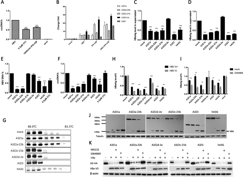
Fig. 1.
The tsAPOBEC3 proteins restrict HBV replication, which can be counteracted by HBx protein. a PTHs (1.6 × 10⁵ cells) infected with HBV (105 copies) and treated with IFN-ɑ (1000 IU/ml) or lamivudine (LAM, 3TC) at 0.5 μM. Total DNA was collected to detect cccDNA levels by qPCR after 72 h. b IFN-ɑ (1000 IU/ml)-treated and HBV (105 copies)-treated PTHs (1.6 × 10⁵ cells). Total RNA was used to detect tsAPOBEC3 levels by qPCR after 12 h. c–f pcDNA3.1-tsA3s-HA and pAAV-HBV 1.3B were cotransfected into Huh7 cells; the supernatant was used to detect HBsAg c and HBeAg d by ELISA; and total DNA was used to detect HBV DNA e and cccDNA f levels via qPCR after 72 h. g pcDNA3.1-tsA3s-HA and pAAV-HBV 1.3B were cotransfected into Huh7 cells with a low ratio of 1:5, and total DNA was used to detect G → A mutation by 3D-PCR after 72 h. h pcDNA3.1-tsA3s-HA and HBV X+/HBV X− were cotransfected into Huh7 cells, and the supernatant was used to detect HBeAg by ELISA after 48 h. i pcDNA3.1-tsA3s-HA and HBV X+ were cotransfected into Huh7 cells, then the cells were treated with GW4869 after 6 h to inhibit exosome biogenesis and secretion, and the supernatant was used to detect HBeAg by ELISA after 48 h. j pcDNA3.1-tsA3s-HA and pcDNA3.1-HBx-V5 were cotransfected into 293T cells at 1:0, 1:1, 1:5, and 1:10, and antibodies were used to detect tsA3 levels by Western blot analysis after 48 h. k pcDNA3.1-tsA3s-HA and pcDNA3.1-HBx-V5 were cotransfected into 293T cells at 1:10, then the cells were treated with GW4869 or MG132 after 6 h, and antibodies were used to detect tsA3 levels by Western blot analysis after 48 h

