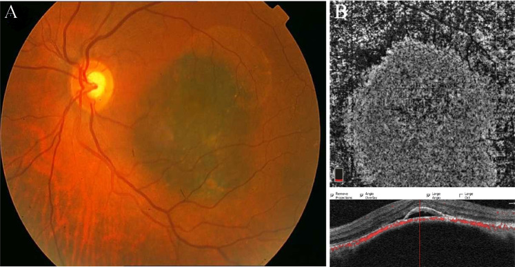
Fig. 2. Choroidal melanoma.
a Fundus photo of the left eye showing an elevated, pigmented lesion in the macula with dispersed orange pigment and subretinal fluid at the inferior margin. b OCTA B-scan and the corresponding en face image demonstrate high vascular flux and the overlaying SRF and pigment epithelial detachment associated with outer retina layer disorganization in the corresponding OCT.

