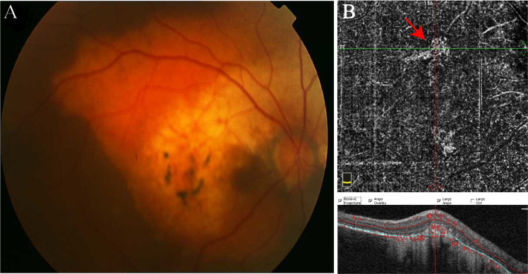
Fig. 7. Choroidal osteoma.
a Fundus photograph represents a circumpapillary amelanotic mass with pseudopod-like projections and areas of ossification. b OCTA depicts quiescent choroidal neovascularization lesion associated with choroidal osteoma (red arrow) at the outer retinal slab. (Colour figure online).

