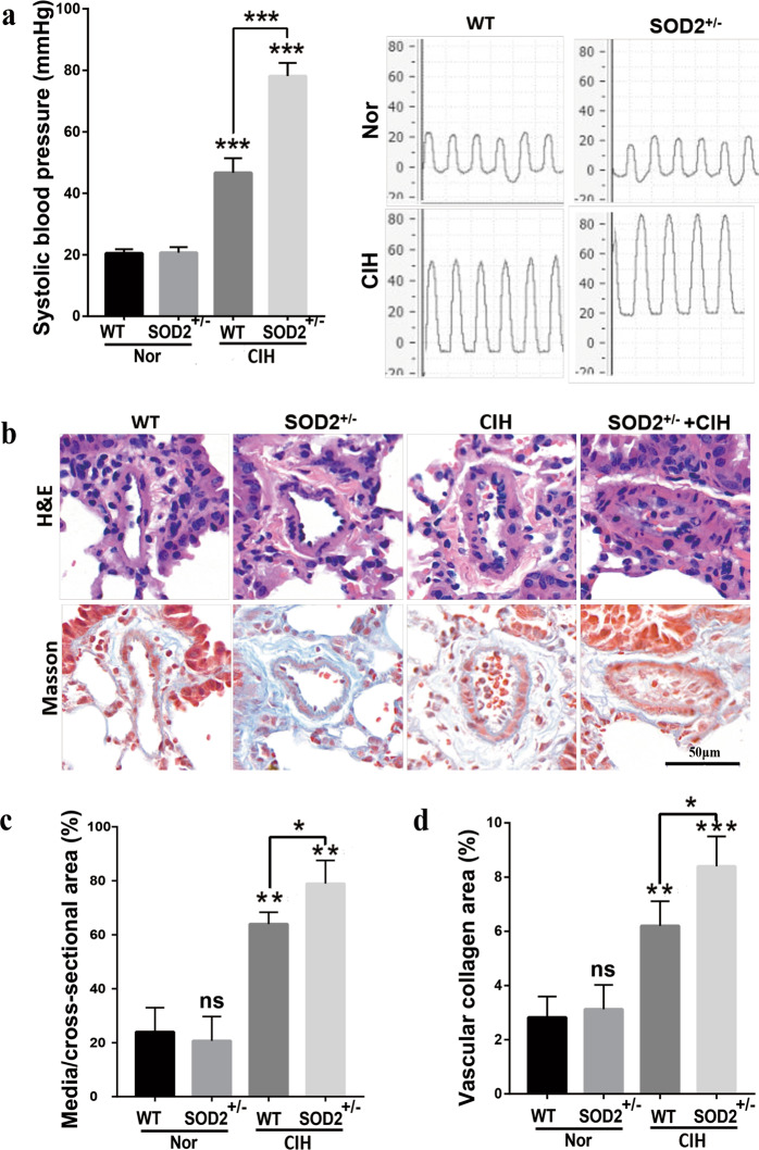
Fig. 2. Deficiency in SOD2 deteriorates CIH-induced pulmonary artery remodeling. WT and SOD2+/− mice were exposed to Nor or CIH conditions for 4 weeks.
a Systolic blood pressure was then measured, and representative photomicrographs are presented. b HE and Masson’s trichrome staining of pulmonary arterioles in the four groups were performed and are presented. c The percentage wall thickness of pulmonary arterioles was defined as the area occupied by the vessel wall divided by the total cross-sectional area of the arteriole. d The vascular collagen area was evaluated by quantitative image analysis of Masson’s trichrome staining. Scale bar: 50 μm. Graph bars represent the mean ± SEM of 20 vessels from 6 mice per group. *P < 0.05, **P < 0.01, and ***P < 0.001 compared with the WT group. Nor normoxic, CIH chronic intermittent hypoxia, HE hematoxylin and eosin, WT wild type.

