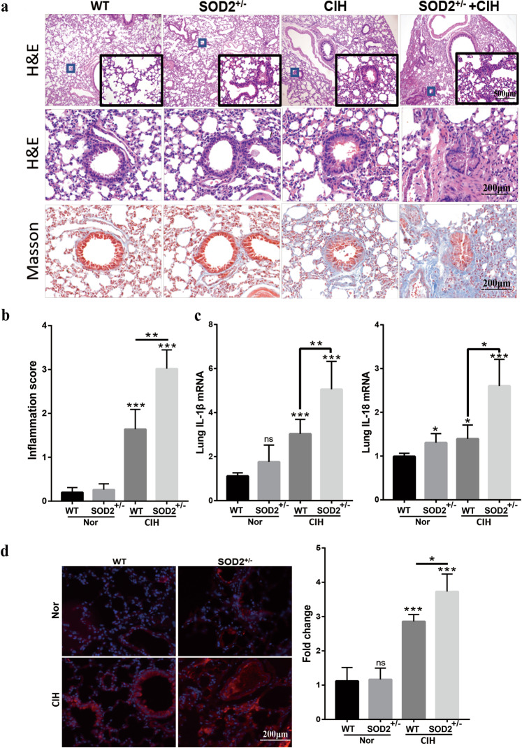
Fig. 3. Deficiency in SOD2 enhances CIH-induced pulmonary inflammation and oxidative stress.
a Morphological observations for the lungs from the WT, SOD2+/−, WT + CIH, and SOD2+/− + CIH groups. Representative light microscopy images of HE and Masson’s trichrome staining are shown. b Pathological scoring results for lung sections from the four groups. c Quantitative reverse transcriptase polymerase chain reaction (RT-PCR) results for the interleukin (IL)-1β and IL-18 mRNA levels in lung lysates. d Immunohistochemical evaluation of ROS in mouse lungs from the four groups. The density of red fluorescence indicates the ROS level. Scale bar: 50 μm. Graph bars represent the mean ± SEM from six mice per group. * P < 0.05, ** P < 0.01, and *** P < 0.001 compared with the WT group. CIH chronic intermittent hypoxia, HE hematoxylin and eosin.

