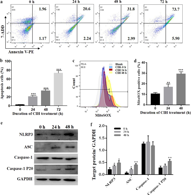
Fig. 5. IH induces apoptosis and mitochondrial oxidative stress in RAW264.7 cells.
a Following IH exposure of RAW264.7 cells for 0, 24, 48 or 72 h, the cells were collected, stained using annexin V-PE/7-ADD and assessed by FACS analysis. b Quantitative analysis of apoptotic cell numbers is shown. c Flow cytometric analysis of cells treated with MitoSOX after IH exposure for 0, 24 or 48 h is shown. d Quantitative analysis of MitoSOX-positive cells is shown. e The protein expression of NLRP3 and ASC was detected by Western blot analysis. f Quantification of relative protein expression was performed by densitometric analysis, and GAPDH acted as an internal control. All data are presented as the mean ± SEM. *P < 0.05, ** P < 0.01 and ***P < 0.001 compared with the control group. Every experiment was repeated at least three times. IH intermittent hypoxia.

