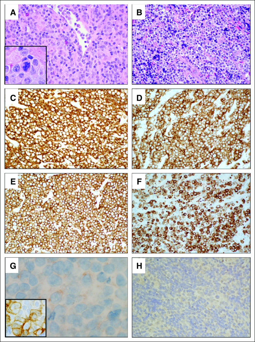
FIG 2.
Lymph node and mediastinal involvement by malignant hematopoietic neoplasm with SMARCB1 deletion showing myeloid, B-cell, and T/natural killer (NK)-cell differentiation. (A) Histologic section of left cervical lymph node revealed diffuse proliferation of atypical mononuclear cells with numerous mitotic figures (inset). There were a few background inflammatory cells (hematoxylin and eosin stain, ×400). (B) Histologic sections of the left anterior mediastinal mass showed atypical mononuclear cells with scattered large cells presenting marked pleomorphism and occasional multinucleation (hematoxylin and eosin stain, ×400). (C) Neoplastic cells showed strong expression of CD45 (leukocyte common antigen; CD45 immunohistochemistry, ×400). (D) CD2 immunohistochemistry showed diffuse and strong expression in neoplastic cells, indicating T/NK-cell differentiation (CD2 immunohistochemistry, ×400). (E) CD7 was diffusely and strongly expressed in neoplastic cells (CD7 immunohistochemistry, ×400). (F) A subset of neoplastic cells shows strong expression of CD79a, indicating B-cell differentiation (CD79a immunohistochemistry, ×400). (G) Weak cytoplasmic expression of myeloperoxidase was seen in a subset of cells, and atypical cells also show immunoreactivity to CD13 immunohistochemistry (inset), supporting myeloid differentiation (myeloperoxidase immunohistochemistry, ×1,000; CD13 immunohistochemistry, ×1,000). (H) Immunohistochemistry for myogenin demonstrated that these cells did not show rhabdoid differentiation (myogenin immunohistochemistry, ×400).

