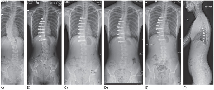
Fig. 2.
(A) An 11-year-old girl with juvenile idiopathic scoliosis Sanders 2. (B) Treated using anterior spinal tethering between T6 and L1. (C) Six-month postoperative radiograph demonstrates growth modulation: L1 is levelled. (D) One-year follow-up shows splaying between T9 and T10 screws. This suggests rupture of the tether. (E) Two-year follow-up shows a stable curve without further progression despite tether rupture. (F) Two-year lateral spinal radiograph demonstrates maintenance of the sagittal balance.

