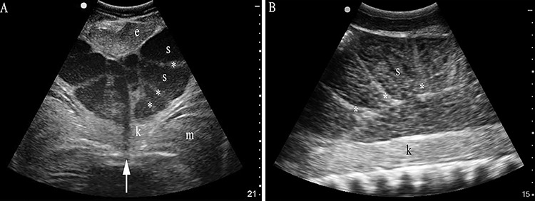
Figure 1.

Semen localization in ampulla epididymis by ultrasonography in spring mating season. With the transducer in transverse orientation (A) and positioned on midline (arrow) or sagittal and positioned laterally (B) immediately cranial to the pelvic girdle, semen (s) in the ampulla lumen is hypoechoic compared to kidney (k), dorsal musculature (m), and the ventral and caudal epigonal (e). Semen is separated by septae (asterisk) forming partial chambers in the terminal portion of the ampulla. Imaging depth (cm) is given on the right of each sonograph.
