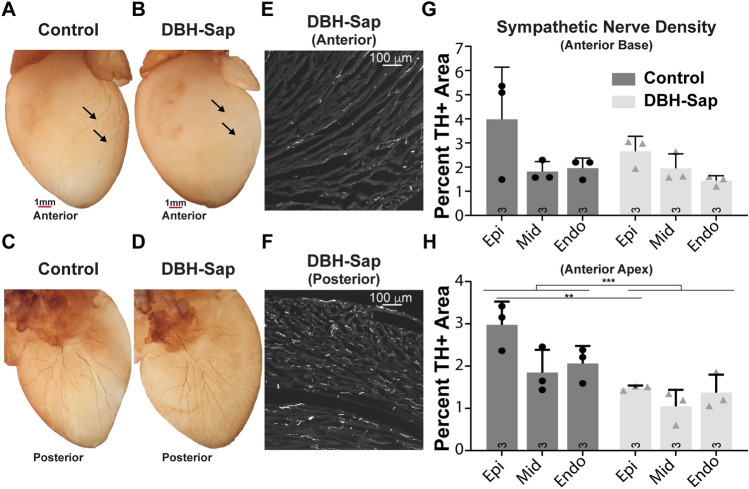
Figure 1.
Effects of regional toxin application on sympathetic nerve fiber density. (A,B) Whole-heart labeling of tyrosine hydroxylase (TH) demonstrates fewer visible sympathetic nerve fibers on the anterior surface (where toxin solution is applied) of the DBH-Sap heart (B) compared to control (A). (C,D) The posterior surface is similar between control and DBH-Sap groups. (E,F) Short axis TH images of the anterior (E) and posterior (F) surface of a DBH-Sap mouse heart. (G,H) Sympathetic nerve fiber density from anterior base (G) and apex (H) regions 5 days after treatment. At the apex, the percent TH + area was significantly reduced in the epicardium of the DBH-Sap group, and there was also a main effect between groups (***p < 0.001, main effect control vs. DBH-Sap, two-way ANOVA, H). Data are mean ± SD; analyzed with GraphPad Prism 8.3 (GraphPad Software, San Diego, CA, USA); control: n = 3; DBH-Sap: n = 3; **p < 0.01, ***p < 0.001.

