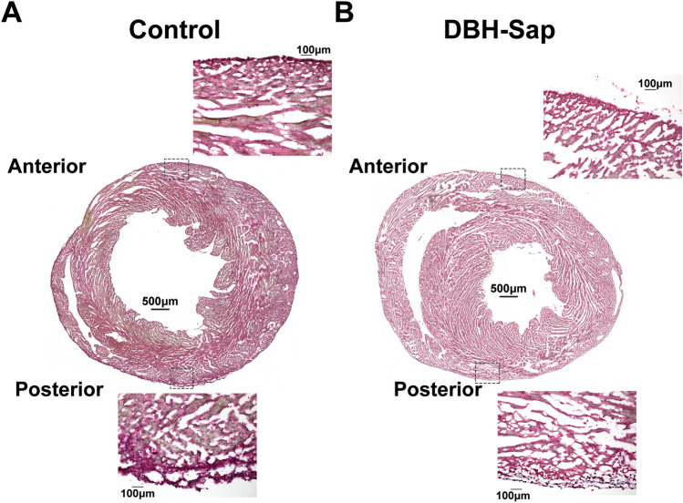
Figure 2.
Masson’s trichrome staining of short-axis sections. (A) Cross-sectional staining of a control heart and 20 × images of the epicardial/sub-epicardial regions from anterior and posterior surfaces of the heart. (B) Cross-sectional staining of a DBH-Sap (toxin-exposed) heart and 20 × images of the epicardial/sub-epicardial regions from anterior and posterior surfaces. No overt myocardial damage was observed in any heart from either group.

