Figure 6.

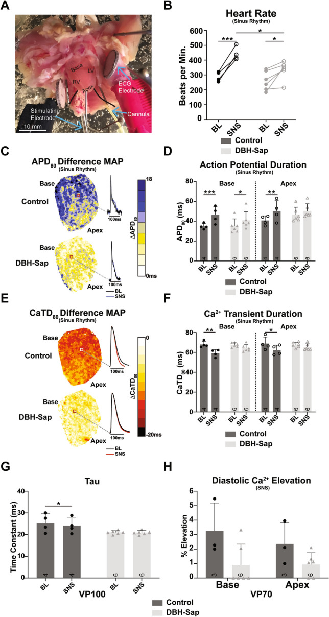
Electrophysiological responses to sympathetic nerve stimulation (SNS) during sinus rhythm and with pacing. (A) Photograph of the mouse innervated heart preparation. (B) Heart rate in control and DBH-Sap groups at baseline (BL) and with SNS. (C) Example difference maps (ΔAPD80) demonstrating APD prolongation with SNS compared to BL. (D) Mean APD80 from base and apex regions at BL and with SNS in both groups. (E) Example difference maps (ΔCaTD80) demonstrating CaTD shortening with SNS compared to BL. (F) Mean CaTD80 from base and apex regions at BL and with SNS in both groups. For panels (B–F) hearts are in sinus rhythm with and without SNS. (G) Time constant of Ca2+ decay (tau) of the whole heart at BL and with SNS in both groups at a pacing cycle length of 100 ms. (H) Mean diastolic Ca2+ elevation at base and apex regions of both groups following a 70 ms pacing train with SNS. Data are mean ± SD; analyzed with GraphPad Prism 8.3 (GraphPad Software, San Diego, CA, USA); control: n = 3–4; DBH-Sap: n = 6; *p < 0.05, **p < 0.01, ***p < 0.001.
