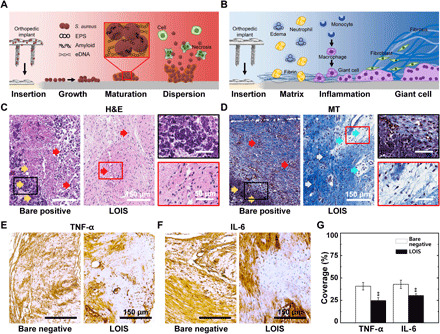
Fig. 5. Histological analysis of tissues surrounding implants incubated in bacterial suspension for 12 hours.

(A) Schematics of the biofilm formation and spreading mechanism on the infected orthopedic implant’s surface. eDNA, extracellular DNAs. (B) Schematics of immune response upon the orthopedic implants’ insertion. (C) H&E staining and (D) MT staining of the tissues around the orthopedic implants of bare positive and LOIS. IHC of immune-related cytokines, (E) TNF-α and (F) IL-6, staining images of the rabbit implanted with bare negative and LOIS. (G) Quantification of the cytokine expression via area coverage measurements (**P < 0.01).
