
Fig. 3. Filia deletion impairs hippocampal NSPC proliferation and neurogenesis.
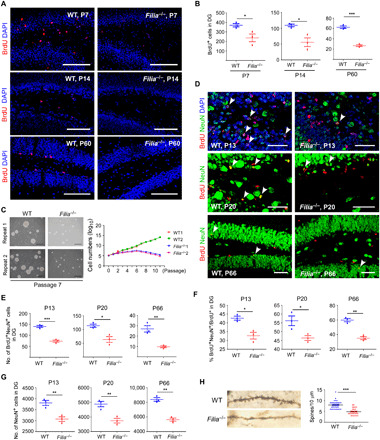
(A) Filia loss compromised hippocampal NSPC proliferation, as monitored by BrdU incorporation when examined at P7, P14, and P60, respectively. Scale bars, 100 μm. (B) Quantification of BrdU+ cells in the dentate gyrus at P7, P14, and P60. (C) NSPCs were derived from the hippocampus of newborn mice and cultured as neurospheres. Left: Photos of neurospheres at passage 7. Right: Growth kinetics of neurospheres from WT and Filia−/− mice. Scale bars, 200 μm. (D) Filia loss impaired the hippocampal neurogenesis in the dentate gyrus. BrdU+NeuN+ cells indicate newborn neurons. Scale bars, 50 μm. (E) Quantification of newborn neurons in the dentate gyrus at P13, P20, and P66. (F) The percentages of NSPCs undergoing neuronal differentiation. (G) Total numbers of neurons in the dentate gyrus. (H) Hippocampal neurons from Filia−/− mice contained much fewer spines than those from WT mice. Left: Morphology of a single neuron from WT and Filia−/− mouse. Right: Spine densities in neurons from WT and Filia−/− mice. Data were represented as means ± SEM; two-tailed Student’s t test, *P < 0.05, **P < 0.01, and ***P < 0.001.