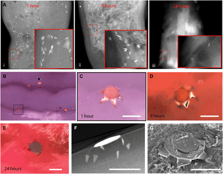
Fig. 4. Theragripper attachment to rat colon and retention upon rectal delivery.

(A) μ-CT images showing the retention of theragrippers in the rat colon (i) 1, (ii) 9, and (iii) 24 hours after rectal administration. The insets show zoomed-in images of the red outlined regions. (B) Postmortem optical examination of the rat colon showing attachment of several theragrippers, and (C) a zoomed-in image of the section shown by dotted lines in (B). (D and E) Optical image showing a theragripper attached to the colon (D) 9 and (E) 24 hours after rectal administration. (F) Postmortem μ-CT image showing the claws of a theragripper latching into the colon luminal surface in vivo. (G) SEM image showing the attachment of the theragripper to the mucosa after rectal administration. Scale bars, 100 μm. (C), (F), and (G) are obtained 1 hour after rectal administration of the theragrippers in the rat.
