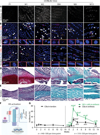
Fig. 1. Ciliogenesis of tendon enthesis cells during postnatal development coincides with Hh signaling and enthesis mineralization.

(A to C) Immunofluorescence staining of Hh component Gli1 (gray) at the supraspinatus tendon enthesis (A) and primary cilia via acetylated tubulin (red, white arrowheads) at the enthesis (B) and midsubstance (C). Panels below (B) and (C) are magnified images corresponding to the colored rectangles. Arrowheads mark primary cilia; 4′,6-diamidino-2-phenylindole (DAPI) stains nuclei; P1, postnatal day 1; W1, postnatal week 1. Scale bars, 10 μm (panel insets). (D and E) Safranin O staining of the tendon enthesis and midsubstance at different developmental stages. Dashed lines mark the tendon enthesis. (F) Illustration of the transduction of Hh signaling inside the primary cilium. IFT, intraflagellar transport protein. (G) Quantification of the incidence of ciliated cells at the tendon and tendon enthesis as well as Gli1-positive (Gli1+) cells, normalized by the total cell number. Four to five mice were analyzed per time point; all data are represented as means ± SD; n represents the number of cells counted. a, P < 0.05 compared to W0; b, P < 0.05 compared to W1; c, P < 0.05 compared to W2; d, P < 0.05 compared to W4; e, P < 0.05 compared to W8; f, P < 0.05 compared to W13.
