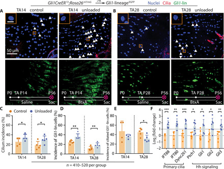
Fig. 2. Unloading during tendon enthesis development stimulates primary cilia assembly.

(A and B) Immunofluorescence for primary cilia (red, white arrowheads) of the tendon enthesis from GliCreERT2;Rosa26mT/mG mice with TA injection and BtxA-induced paralysis (unloaded, right shoulder) or saline injection (control, left shoulder) at the time points indicated. Panels on the top left show magnified views of the orange rectangles. DAPI stains nuclei; P0, postnatal day 1; TA, tamoxifen injection; Sac, sacrifice; scale bar, 10 μm (inset). (C) Incidences of ciliated cells per all the cells at the tendon enthesis of (A) and (B) mice with TA injection at P14 and P28. n represents the number of cells counted. The error bars show SD of tissue samples from different animals. *P < 0.05, **P < 0.01, ***P < 0.001. (D) Incidences of Gli1-lin cells per all the cells at the tendon enthesis of (A) and (B) mice. (E) Incidences of ciliated Gli1-lin cells normalized by Gli1-lin cells at the tendon enthesis, showing the incidence of Gli1-lin cells that were ciliated. (F) Expression of genes related to primary cilia and Hh signaling from tendon entheses of 8-week-old C57BL/6J mice paralyzed since birth.
