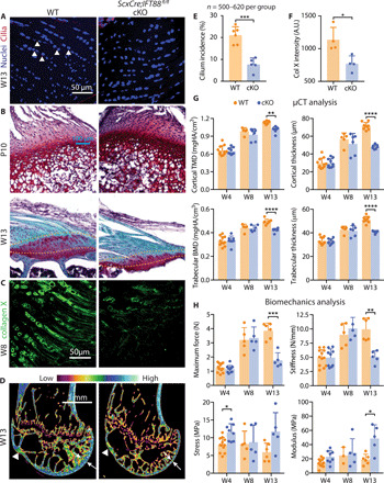
Fig. 4. Primary cilia are necessary for in tendon enthesis formation.

(A) Immunofluorescence for primary cilia (red, white arrowheads) of tendon entheses from 13-week-old ScxCre;IFT88fl/fl (cKO) mice and their WT littermates. (B) Safranin O staining of the tendon entheses from WT and cKO mice at different time points. Dashed lines mark the tendon enthesis. (C) Immunostaining of collagen X at the tendon entheses from 8-week-old cKO and WT mice. (D) Microcomputed tomography (μCT) sections of humeral heads from 13-week-old WT and cKO mice. White arrowheads denote the supraspinatus tendon enthesis; white arrows denote bone thickness of humerus head. Color scale indicates low-to-high bone density. (E) Incidence of ciliated cells from (A). (F) Fluorescent intensity of collagen X (Col X) at the tendon enthesis from 8-week-old cKO and WT mice [e.g., as shown in (C)]. *P < 0.05, **P < 0.01, ***P < 0.001, ****P < 0.0001. A.U., arbitrary units. (G) Humeral head bone morphometry from 4-, 8-, and 13-week-old WT and cKO mice. TMD, tissue mineral density; BMD, bone mineral density. (H) Mechanical properties of the tendon enthesis from 4-, 8-, and 13-week-old WT and cKO mice.
