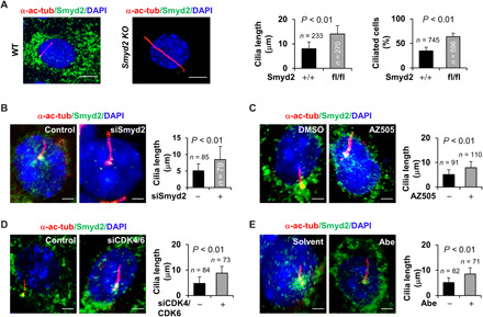
Fig. 3. Depletion of Smyd2 promotes cilia assembly in mouse renal epithelial cells.

(A) Immunofluorescence staining of primary cilia with α-acetyl-tubulin antibody (α-ac-tubulin) in primary renal epithelial cells isolated from kidneys of wild-type (WT) and Smyd2flox/flox:Ksp-Cre mice, in which Smyd2 was specifically knocked out (KO) by the kidney-specific promoter (Ksp)–driven Cre recombinase in renal epithelial cells and cultured in serum-free medium for 72 hours before subjected to staining. The percentage of ciliated cells and cilia length was measured and statistically analyzed. Scale bar, 5 μm. Error bars represent the SD. N values represent the numbers of cilia (left graph) and cells (right graph), respectively, for each group. (B to E) Knockdown of Smyd2 with siRNA (B) or inhibition of Smyd2 with AZ505 (C) as well as knockdown of CDK4/CDK6 with siRNAs (D) or inhibition of CDK4/CDK6 with Abe (E) in mIMCD3 cells resulted in longer cilia compared to control cells as examined by immunofluorescence with α-acetyl-tubulin and SMYD2 antibodies. Scale bar, 5 μm. Error bars represent the SD. N values represent cilia numbers for each group.
