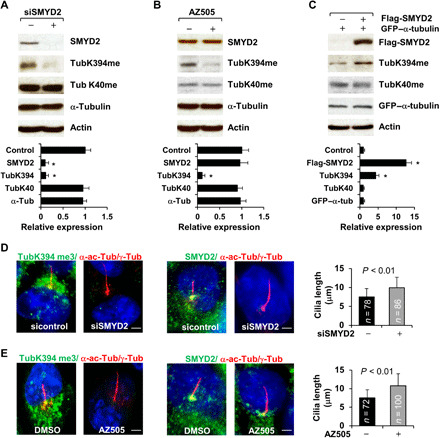
Fig. 5. SMYD2 methylates α-tubulin at lysine-394 (K394) but not at K40.

(A and B) Knockdown of SMYD2 with siRNA (A) or inhibition of SMYD2 with AZ505 (B) decreased methylation of α-tubulin at K394 but not at K40 as examined by Western blotting with our generated TubK40me3 and TubK394me3 antibodies in RCTE cells. The quantification and statistical analysis (n = 3) were shown in the graphs (bottom), in which the band density of control siRNA (A) and DMSO (B) was set to 1. (C) Western blot of the methylation of α-tubulin at K40 and at K394 with the TubK40me3 and TubK394me3 antibodies in RCTE cells transfected with Flag-tagged SMYD2 and GFP-tagged α-tubulin. The quantification and statistical analysis (n = 3) were shown in the graph (bottom). (D) Representative images of RCTE cells stained with TubK394me3 (left) or SMYD2 (right) and costained with α-acetyl-tubulin/γ-tubulin (red) antibodies and DAPI (blue). Scale bar, 5 μm. (E) Representative images of RCTE cells stained with TubK394me3 (left, green) or SMYD2 (right, green) and costained with α-acetyl-tubulin/γ-tubulin (red) and DAPI (blue). Scale bar, 5 μm. Error bars represent the SD. N values represent cilia numbers in (D) and (E).
