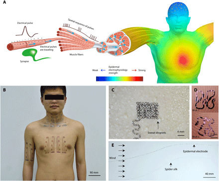
Fig. 1. Illustration of epidermal electrophysiology at scale.

(A) Schematics of spatially varying electrophysiology over skin surface originating from internal muscle and organ activities. (B) Photograph of the body-scale epidermal electrodes. Photo credit: Youhua Wang, State Key Laboratory of Digital Manufacturing Equipment and Technology, Huazhong University of Science and Technology and Flexible Electronics Research Center, Huazhong University of Science and Technology. (C) Open-mesh filamentary serpentine network is unobstructive to sweating and sweat evaporation. Photo credit: Chao Hou, State Key Laboratory of Digital Manufacturing Equipment and Technology, Huazhong University of Science and Technology and Flexible Electronics Research Center, Huazhong University of Science and Technology. (D) Optical micrographs of two filamentary serpentine ribbons conforming to fingerprint topologies. Photo credit: Lin Xiao, State Key Laboratory of Digital Manufacturing Equipment and Technology, Huazhong University of Science and Technology and Flexible Electronics Research Center, Huazhong University of Science and Technology. (E) A filamentary serpentine ribbon flutters in wind similar to a single strand of spider silk. Photo credit: Chao Hou, State Key Laboratory of Digital Manufacturing Equipment and Technology, Huazhong University of Science and Technology and Flexible Electronics Research Center, Huazhong University of Science and Technology.
