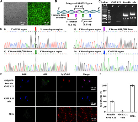
Fig. 5. Analysis of K562 3.21 cells for HBB/GFP gene integration into the AAVS1 site.

(A) Fluorescence microscopy images of purified HBB/GFP-knockin K562 3.21 cells after 20 rounds of culture expansion. (B) Schematic depicting the integration of HBB/GFP gene into the AAVS1 site, in which the locations of the two HDR junctions and four genome-donor boundaries are labeled. (C) Electrophoretogram shows the two characteristic DNA fragments, i.e., the 5′ junction (1.1 kb) and the 3′ junction (1.2 kb). (D) Sanger sequencing of the four genome-donor boundaries. (E) Representative IF images of HBB/GFP-knockin K562 3.21, K562 3.21, and RBCs. (F) qPCR for quantification of HBB mRNA expression.
