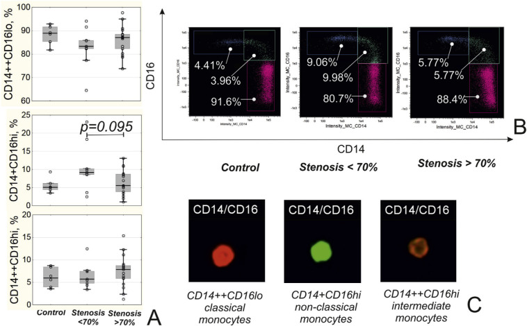
Fig. 1.
Subpopulations of monocytes in patients depending on the presence of coronary stenosis. A) Level of different populations of monocytes in patients. B) Representative dot plots of different monocytes subpopulations. C) Representative images of classical (CD14++CD16lo), non-classical (CD14+CD16hi) and intermediate (CD14++CD16hi) monocytes obtained during the imaging flow cytometry.

