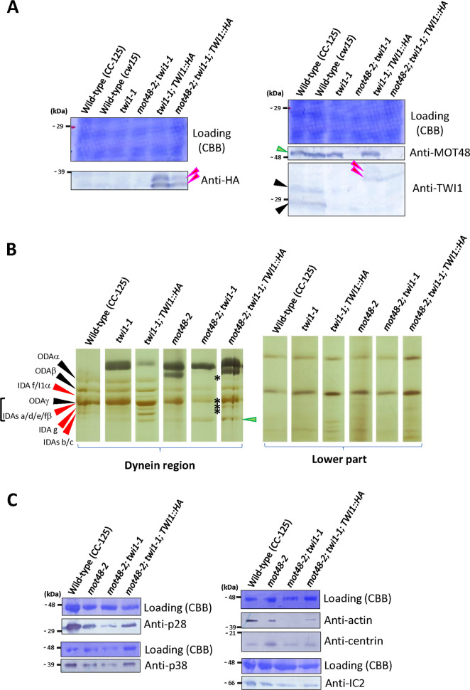
Fig 5. Exogeneous TWI1 protein can rescue the Twi1 phenotype.
A) Immunoblot of whole cell samples from wild-type (CC-125 and cw15), twi1-1, mot48-2; twi1-1, twi1-1; TWI1::HA and mot48-2; twi1-1; TWI1::HA strains using anti-HA (left)/MOT48 and TWI1 (right) antibodies. The black arrowheads indicate the wild-type endogenous TWI1 protein. Red arrowheads indicate the exogeneous TWI1 protein with the 3HA tag. The cDNA-driven exogeneous TWI1::3HA proteins show two bands in these blots (red arrowheads). A green arrowhead indicates the MOT48 protein. B) Urea-PAGE of axonemes from wild-type (CC-125), twi1-1, twi1-1; TWI1::HA, mot48-2, mot48-2; twi1-1, and mot48-2; twi1-1; TWI1::HA strains. For presentation, gel regions of ciliary dyneins and lower parts are shown. The relative positions of ciliary dyneins were adjusted between all strains for comparison. The black arrowheads indicate the HCs of ODA. The red arrowheads indicate the HCs of IDAs. HCs of ODAγ and IDAs a, d, e and fβ form a large band in the urea gel. A green arrowhead indicates HC degradation products. In the mot48-2; twi1-1 strain, the ODAα and IDA bands were missing (asterisks), but these dyneins were recovered in the mot48-2; twi1-1; TWI1::HA strain. The correspondence between bands in the Urea-PAGE gel and DHCs was based on [65–67]. C) Immunoblots of axonemal samples from wild-type (CC-125), mot48-2, mot48-2; twi1-1 and mot48-2; twi1-1; TWI1::HA strains using dynein-subunit antibodies (anti-p28/IDA4, p38, actin/IDA5, centrin/VFL2, and IC2/IC69/ODA6; S1 Table).

