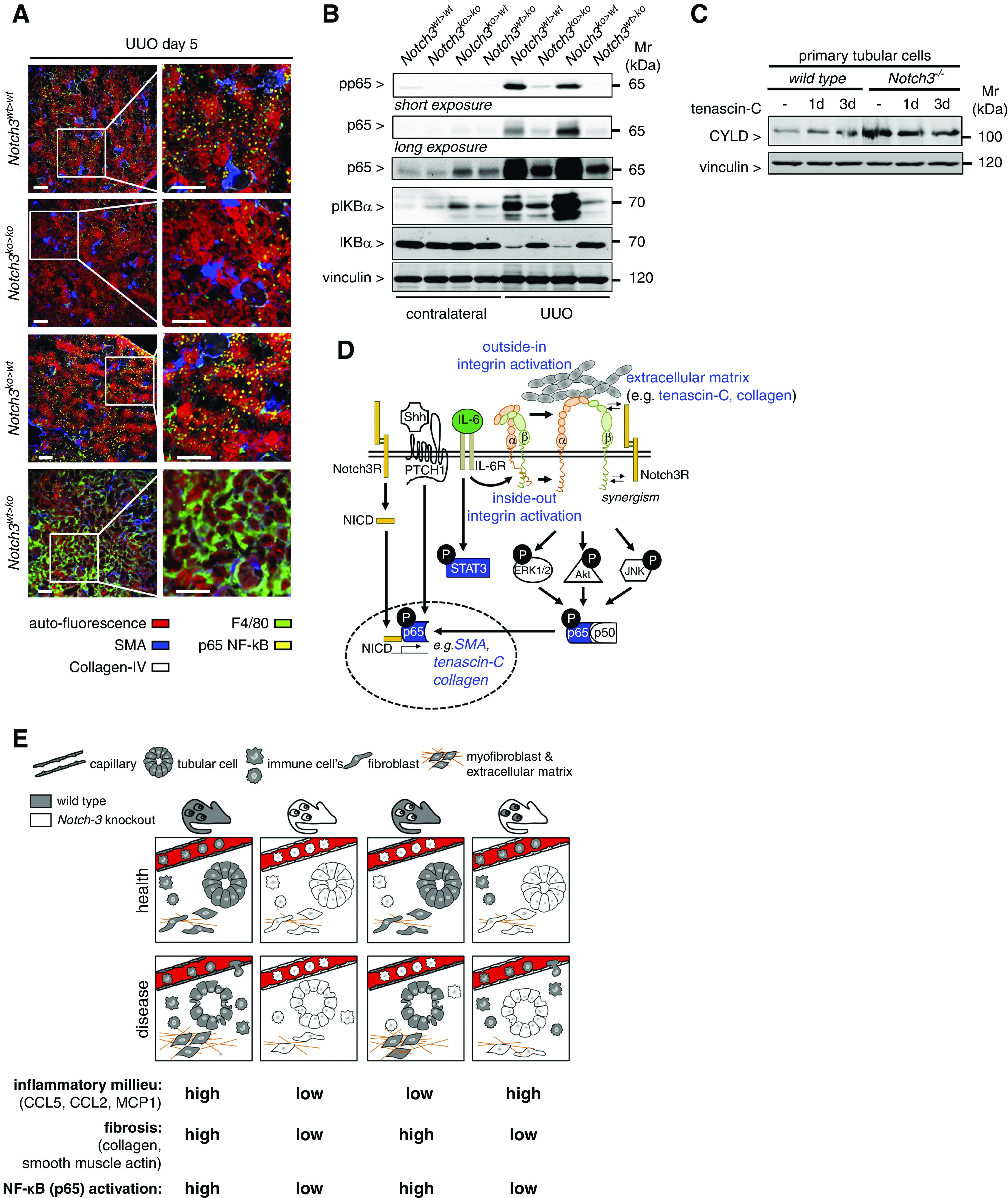
Figure 8.

NF-κB (p65) activation in diseased kidneys is dependent on Notch3 expression on tissue-resident cells. (A) Representative images obtained by automated multidimensional fluorescence microscopy (MELC) visualize the spatial distribution of NF-κB–positive cells in diseased kidneys from the four different chimeric animal strains (NF-κB p65 is stained in yellow). F4/80 in green identifies immune cells. Collagen type IV (white) and SMA (blue) allow for discrimination of tissue structures, tubular cells exhibit a red autofluorescence signal. n=3. Scale bars, 50 μm. (B) Western blot analyses were performed with kidney tissue lysates. Antibodies specific for NF-κB (p65), phospho (Ser536)–NF-κB (p65) (with long exposure to visualize the signals in the contralateral kidneys), total IκBα, and phospho (Ser32)–IκBα were applied. (C) Western blot analysis of whole-cell lysates from TNC-stimulated primary tubular cells (WT or Notch3 knockout) revealed an upregulation of the NF-κB inhibitor CYLD in Notch3-knockout cells. Experiments were repeated three times. (D) Schematic illustrating the crosstalk of receptor Notch3 in combination with Shh and integrin signaling leading to the accumulation of ECM. All data obtained within this study are highlighted in blue. Known interactions from the literature are indicated as black lines with white boxes. (E) Cartoon that depicts key findings of the study.
