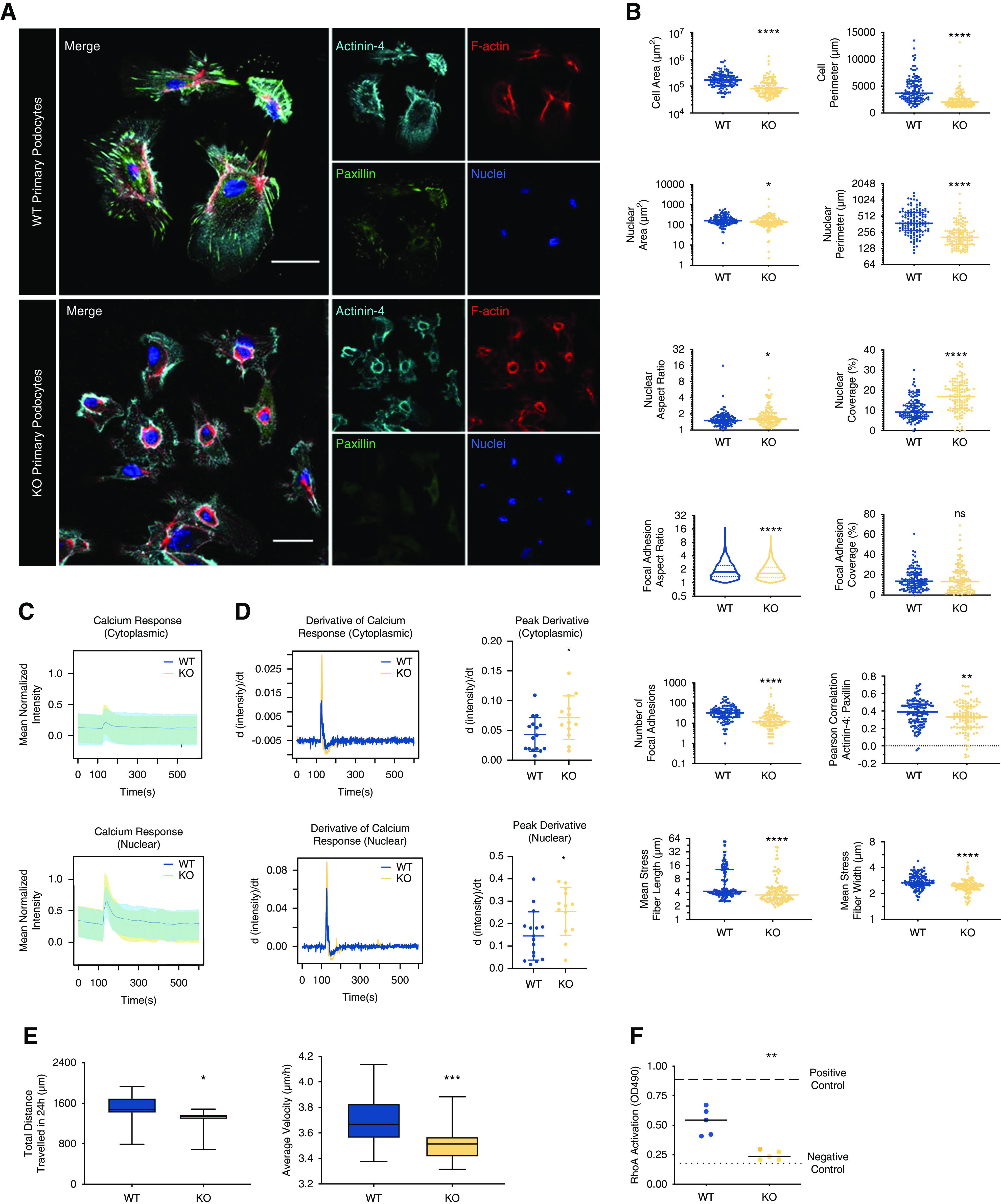
Figure 3.

LIM-nebulette plays key cell biologic roles in primary mouse podocytes. (A) Representative confocal immunofluorescence images of primary mouse podocytes isolated from WT or Nebl−/− mice (KO) stained for focal adhesion marker paxillin (green), actin crosslinker actinin-4 (cyan), F-actin (red), and nuclei (blue; scale bars, 50 μm). (B) HCA shows that KO cells (n=121) have significantly different focal adhesion, cellular, nuclear, and cytoskeletal morphometrics including smaller spreading area and nuclei, fewer and shorter focal adhesions, and thinner and shorter stress fibers (*P<0.05, **P<0.01, ****P<0.001; nonparametric unpaired t test, nWT=121, nKO=120). Live-cell imaging using Fluo-4 calcium sensor shows that both (C) the amplitude and (D) the rate of transient uptake of calcium (derivative of intensity) are significantly affected in KO cells (*P<0.05; nonparametric unpaired t test, n=13–16 cells for each independent experiment). Live-cell imaging also shows that basal cell motility is significantly altered in KO cells. (E) Total distance traversed in 24 hours, and average basal cellular velocity (*P<0.05, ***P<0.001; nonparametric unpaired t test, n>900 cells in each group). (F) Average RhoA GTPase activity levels, as measured by G-LISA, of freshly isolated and flash-frozen glomeruli from WT or KO mice show significant reduction in RhoA activity in KO podocytes (**P<0.01; repeated measures ANOVA, n=6).
