
Figure 5.
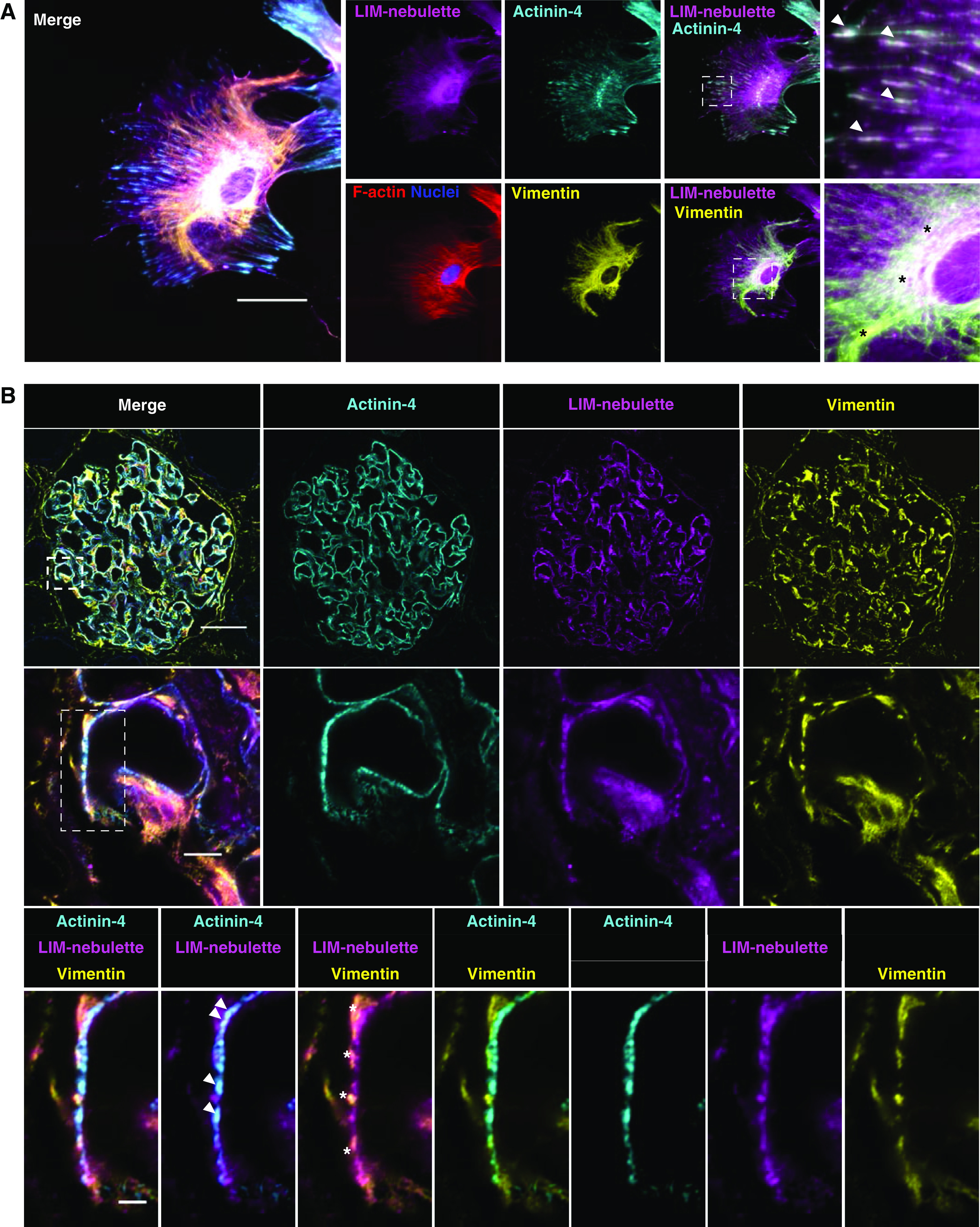
LIM-nebulette colocalizes with focal adhesions as well as intermediate filaments in human podocytes in vitro and in vivo. (A) Representative five-color immunofluorescence images showing that substantial LIM-nebulette (magenta) expression was detected in hiPSC-derived podocytes observed as punctate in the cell periphery and filamentous clusters in the perinuclear core. Arrows point to peripheral expression of LIM-nebulette that colocalizes with actinin-4 (cyan), whereas asterisks point to perinuclear expression of LIM-nebulette colocalizing with vimentin (yellow) intermediate filaments (scale bar, 50 μm). (B) This was recapitulated in human podocytes in situ; representative immunofluorescence images of healthy human glomerulus taken with Zeiss Airyscan super-resolution confocal microscope (top to bottom scale bars for each magnification are 50, 5, and 2 μm, respectively; arrows highlight LIM-nebulette colocalization with actinin-4, whereas asterisks point to LIM-nebulette colocalization with vimentin).