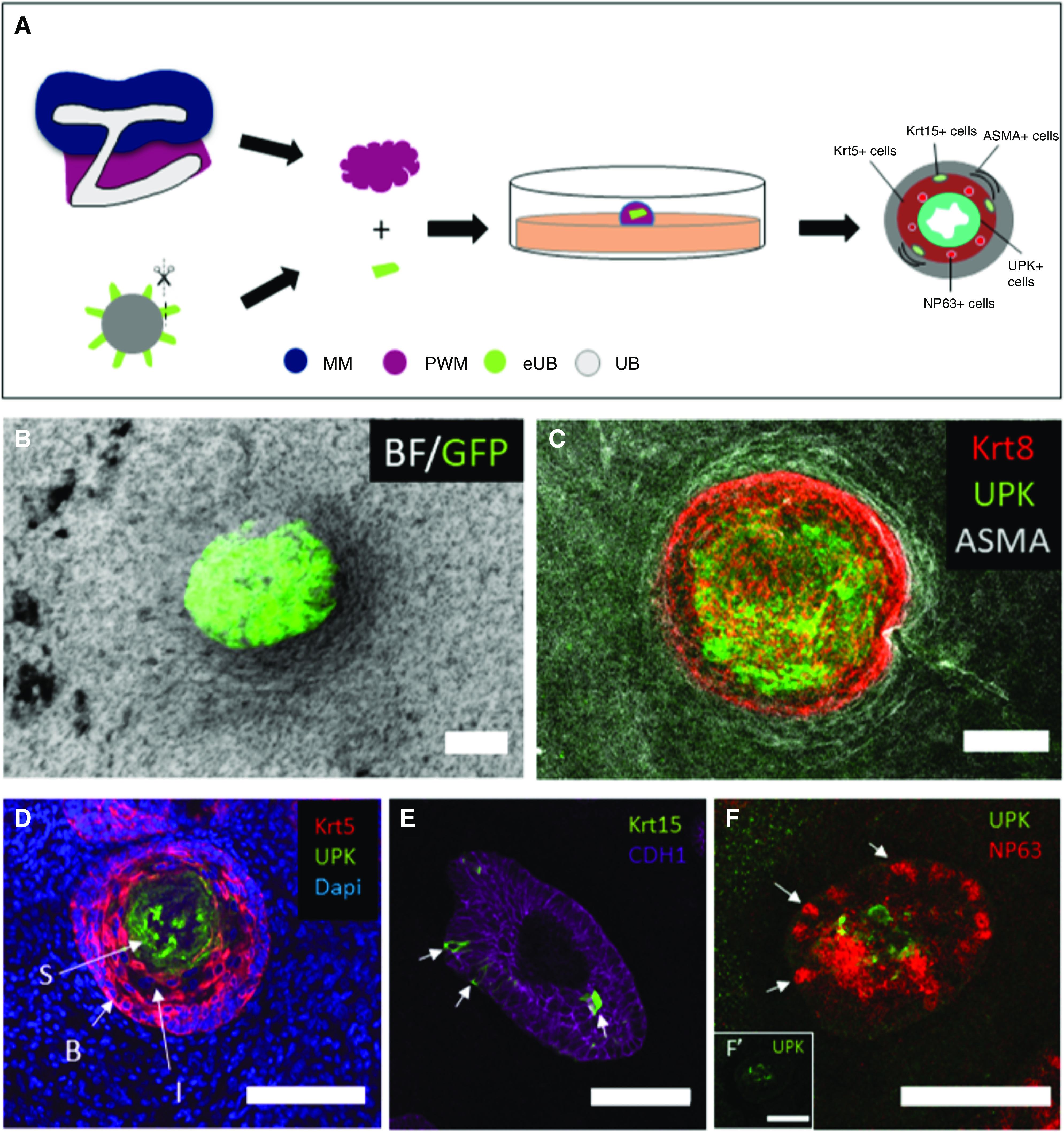
Figure 4.

Urothelial differentiation in pure peri-Wolffian mesenchyme. (A) Steps of recombination of Hoxb7-GFP-eUB with PWM cells. (B) A combined bright-field (BF) and GFP image of a Hoxb7-GFP eUB recombined with PWM cells. (C) Immunofluorescence stain of an eUB recombined with PWM cells showing expression of UPK in the adluminal epithelium, KRT8 in the urothelium as a whole, and smooth muscle actin (ASMA) around the epithelium. (D) A 6-μm section of an eUB combined with PWM shows UPKIII expression in superficial (“S”) cells, KRT5 in basal (“B”) cells, and also the presence of KRT5- intermediate (“I”) cells in the less basal zone of the area otherwise dominated by B cells. (E) Krt15 is expressed by occasional cells in the B cell layer; the counterstain E-cadherin (Cdh1) marks all epithelial cells of the eUB graft. (F) Cells expressing the intermediate cell marker NP63 (arrows) and others expressing UPK; the insert (F') shows the UPK channel alone, for clarity. MM, metanephric mesenchyme; PWM, peri-Wolffian mesenchyme.
