Figure 6.

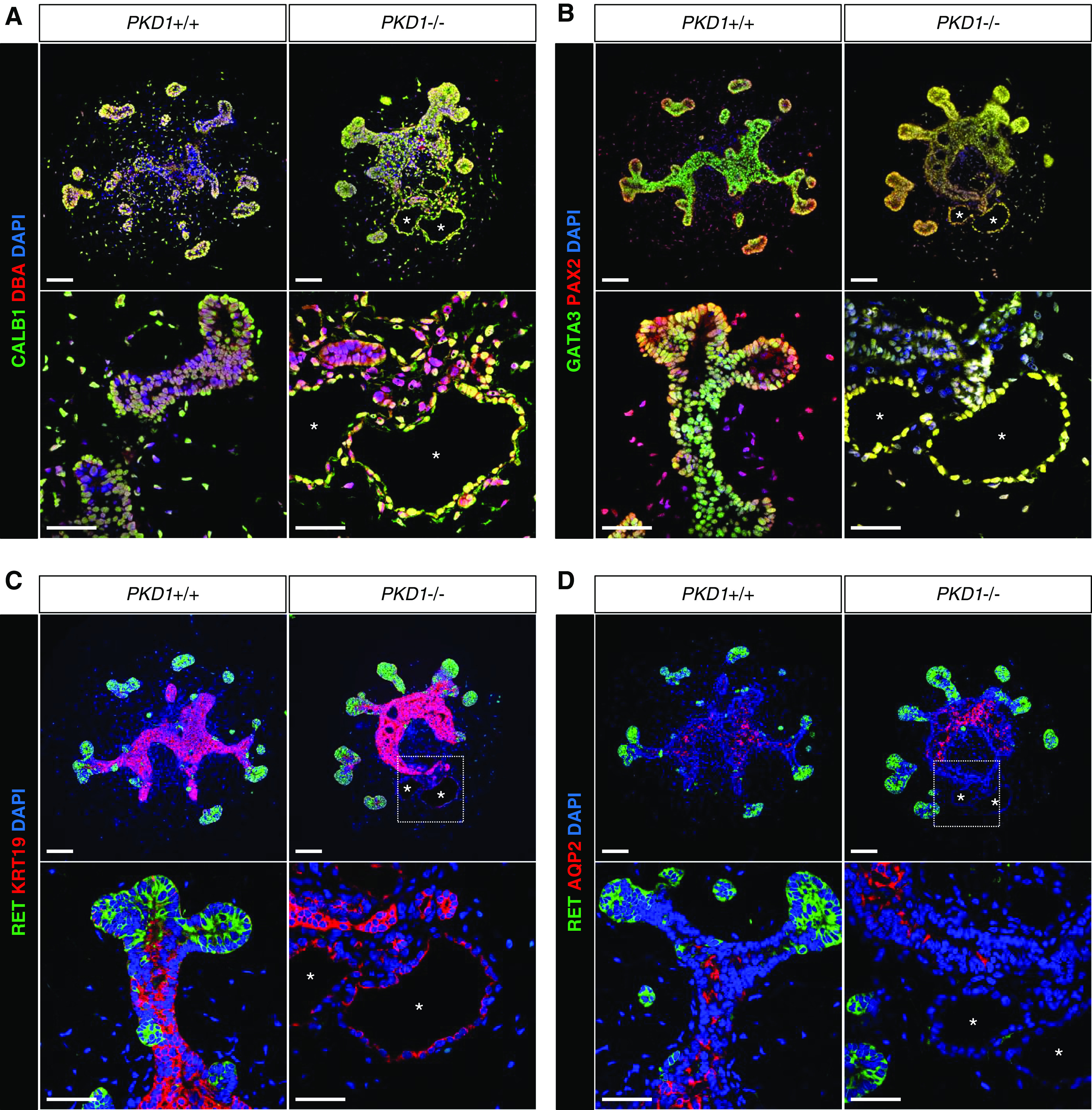
PKD1 mutant cysts express markers for UB epithelia. Confocal images of immunofluorescence in UB organoids at day 24. (A) CALB1 (green) and DBA (red). (B) GATA3 (green) and PAX2 (red). (C) RET (green) and KRT19 (red). (D) RET (green) and AQP2 (red). The asterisks indicate the lumens of cysts. Scale bars, 100 μm (upper panels) and 50 μm (lower panels). DAPI, 4′,6-diamidino-2-phenylindole.
