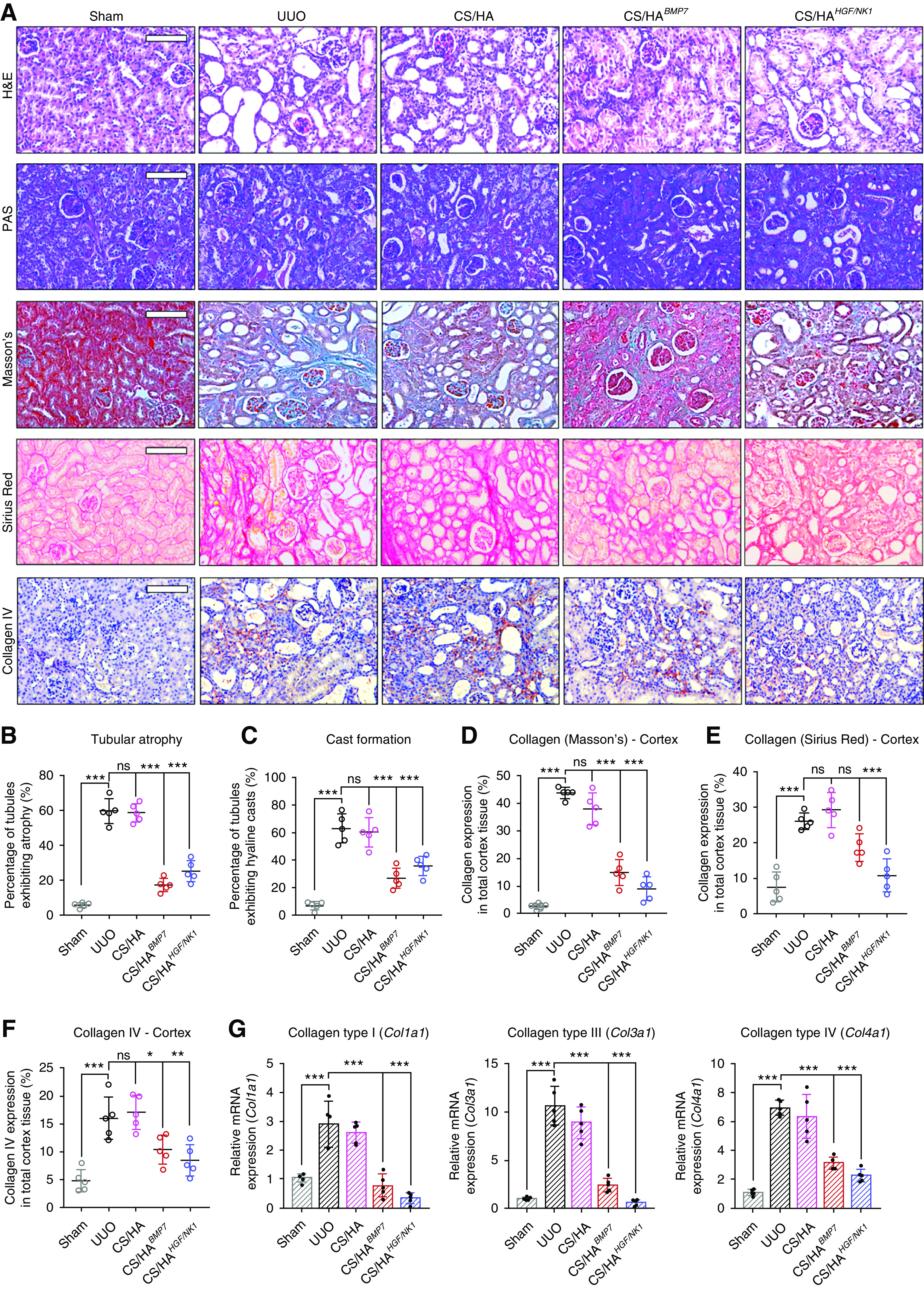
Figure 5.

CS/HA-mediated overexpression of BMP7 or HGF/NK1 prevents in vivo development of CKD pathology. (A) Histologic staining of kidney sections 7 days post UUO surgery (n=5). Top row, H&E; second row, PAS; third row, Masson’s trichrome; fourth row, Sirius Red; bottom row, collagen IV immunostaining. Scale bars, 100 µm. (B) Calculated percentage of tubular atrophy and (C) hyaline cast formation, as calculated from averaged three fields of view of cortex images (n=5). (D) Collagen expression in cortex sections as visualized by Masson’s trichrome was quantified by calculating the areas positive for staining (blue) per three separate fields of view (n=5). (E) Collagen expression in cortex sections as visualized by Sirius Red was quantified by calculating the areas positive for staining (red) per three separate fields of view (n=5). (F) Collagen IV expression in cortex sections as visualized by anti-collagen IV immunostaining was quantified by calculating the areas positive for staining (red-brown) per three separate fields of view (n=5). (G) Collagen genes were assessed by qRT-PCR: Col1a1, Col3a1, Col4a1 (n=5). Data are shown as mean±SD, and statistical significance is indicated by: *P≤0.05; **P≤0.01; ***P≤0.001.
