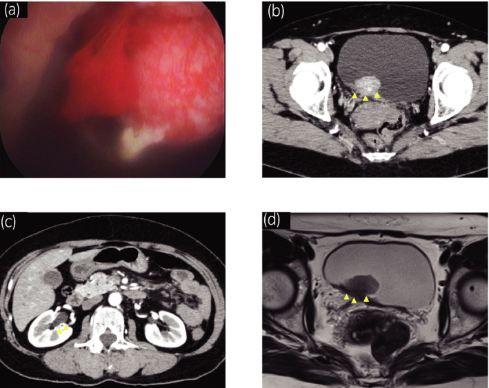
Fig. 1.

Clinical and radiological findings before TURBT. (a) Cystoscopy showed a sessile non‐papillary bladder tumor adjacent to the right ureteric orifice. (b) Abdominal CT with contrast showed a 20‐mm bladder tumor at the right posterior bladder wall and (c) Grade 1 right hydronephrosis. (d) Abdominal MRI showed a 20‐mm bladder tumor suspected for invasion to the muscular layer at the right posterior bladder wall on T2 weighted image.
