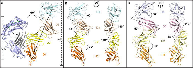
Fig. 1.
Overall structure of HLA-G1-bound LILRB1 and ligand-free LILRB2. a Geometry of the LILRB1 interaction with HLA-G1. The structure of LILRB1 D1 is shown in orange, D2 in yellow, D3 in wheat, and D4 in pale cyan. Light blue indicates the heavy chain of HLA-G1, light green indicates the presented peptide, and gray indicates β2m. b Cartoon backbone representation of LILRB1 displaying the angles between adjacent domains, which are marked the same as in a. c Cartoon backbone representation of LILRB2 displaying the angles between adjacent domains. The structure of LILRB2 D1 is shown in bright orange, D2 in pale yellow, D3 in light pink, and D4 in light blue

