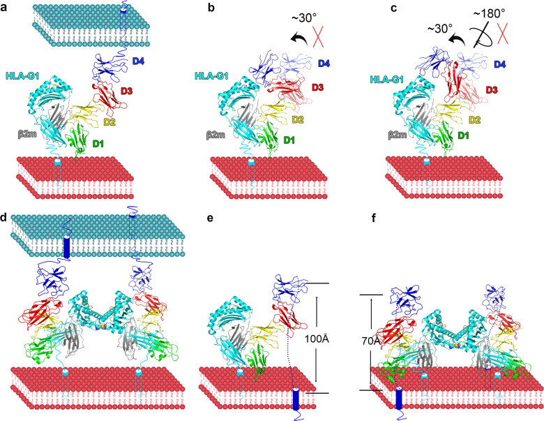
Fig. 6.
Models of full-length LILR binding to HLA-I in cis and trans. The green, yellow, red, and blue colors indicate the four domains in LILRB1/2. The cyan, gray, and magenta represent the HLA heavy chain, β2m, and peptide, respectively. a Trans interaction between LILRB1/2 and HLA-I. b The proposed interaction mode in which D3 and D4 bend to interact with HLA-I and the peptide. c The proposed interaction mode in which the acute interchain angle between D3 and D4 faces the HLA-I and peptide, allowing the interaction between D3D4 and the peptide as well as HLA-I. In b and c, D3D4 in the complex structure between LILRB1 and HLA-G1 are displayed at 50% transparency, while those with no transparency indicate D3D4 in the two proposed models.25,26 d Trans interaction between LILRB1/2 and an HLA-G1 dimer. e Cis interaction between LILRB1/2 and HLA-I. f Cis interaction between LILRB1/2 and an HLA-G1 dimer

