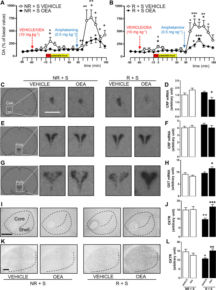
Fig. 3. OEA treatment dampened AcbSh DA release induced by stress exposure or amphetamine challenge. OEA treatment affected CRF, oxytocin mRNA levels, and oxytocin receptor expression in bingeing rats.
Time course of extracellular DA levels (expressed as % of basal values) measured in the nucleus accumbens shell of NR + S (non restricted + stressed, a, N = 9–11) and R + S (restricted + stressed, b, N = 6–9) rats during microdialysis experiment. The first three samples were collected before treating rats with vehicle (veh) or OEA (10 mg kg−1, i.p.) and used as baseline (NR + S baseline = 225.5 ± 43.66; R + S baseline = 205.1 ± 21.07, no statistically significant difference); 45 min after treatment, rats were subjected to the stress procedure for 15 min and subsequently received the HPF for 60 min. Thirty minutes after the end of HPF exposure, rats were administered with amphetamine (0.5 mg kg−1, subcutaneous injection (s.c.)). Data are expressed as mean ± SEM. *P < 0.05; **P < 0.01; ***P < 0.001 vs the mean of the first three samples (basal values) within the same group (Dunnett’s multiple-comparison test). °P < 0.05; °°P < 0.01; °°°P < 0.001 vs OEA-treated rats in the same time point of the same diet regimen group (Bonferroni’s test for between-group comparisons). Red arrow: veh or OEA (10 mg kg−1, i.p.) administration; blue arrow: amphetamine administration (0.5 mg kg−1, s.c.). Representative in situ hybridization images (scale bar = 1 mm) of CRF mRNA expression within the central amygdala (CeA, c), CRF, and oxytocin mRNA in the paraventricular nucleus (PVN, e, g) of NR + S (non restricted + stressed) and R + S (restricted + stressed) rats treated with either vehicle (veh) or OEA (10 mg kg−1, i.p.), and sacrificed 60 min after the treatment. Semiquantitative densitometric analyses of CRF mRNA in the CeA (d), and CRF and oxytocin mRNA in the PVN (f, h, respectively) of NR + S and R + S rats treated with either veh or OEA (10 mg kg−1, i.p.), and sacrificed 60 min after treatment. Data are expressed as mean ± SEM. *P < 0.05 vs veh in the same diet regimen group (Tukey’s post hoc test, N = 4–6). Representative photomicrographs (scale bar = 500 μm) showing oxytocin receptor (OXTR) immunostaining within the nucleus accumbens (core and shell, i) and the caudate putamen (k) in brain slices collected from both NR + S (non restricted + stressed) and R + S (restricted + stressed) rats treated with either vehicle (veh) or OEA (10 mg kg−1, i.p.) and sacrificed 120 min after treatment. Semiquantitative densitometric analysis of oxytocin receptor expression within the nucleus accumbens (j) and caudate putamen (l) of NR + S and R + S rats treated with either veh or OEA (10 mg kg−1, i.p.) and sacrificed 120 min after treatment. Data are expressed as mean ± SEM. **P < 0.01; ***P < 0.001 vs veh in the same diet regimen group; °P < 0.05; °°P < 0.01 vs NR + S in the same treatment group (Tukey’s post hoc test, N = 3).

