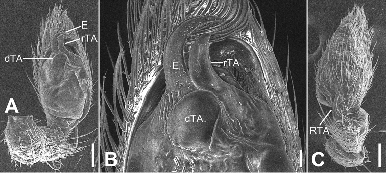
Figure 19.
SEM micrographs of Otacilia zaoshiica sp. nov., palp of male holotype. A ventral view B same, detail of embolus, distal tegular apophysis and retrolateral tegular apophysis C dorsal view. Scale bars: 0.1 mm (A, C), 20 µm (B). Abbreviations: dTA – distal tegular apophysis, E – embolus, RTA – retrolateral tibial apophysis, rTA – retrolateral tegular apophysis.

