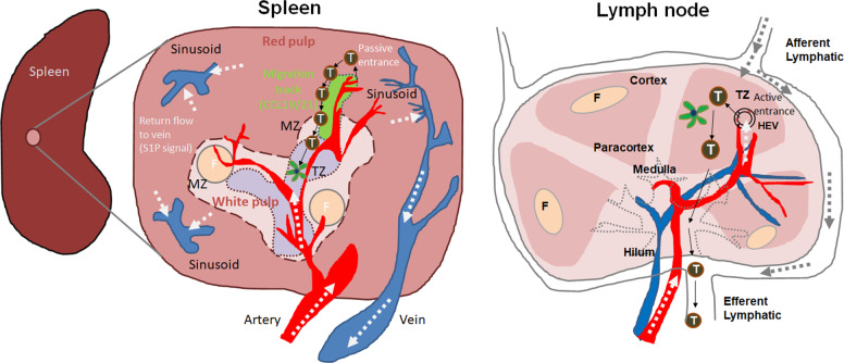
Fig. 1.
T cell trafficking into the spleen versus the lymph nodes. The spleen is largely composed of red and white pulp. T and B cells are concentrated in the white pulp, while blood particulates and red blood cells are filtered in the red pulp. Between the red and white pulp, marginal zones (MZs) are present and contain MZ B cells and macrophages to allow rapid antibody and phagocytic responses, respectively. T cells passively enter the spleen through arterioles in the red pulp and the MZ. Around the arterioles, perivascular stroma cells are present (depicted in green), forming a unidirectional migration track to T zones (TZ) in the white pulp. These cells express CCL19 and CCL21, which chemotactically guide T cells to enter the TZ through MZ bridging channels. The red pulp migration track is connected to white pulp TZ stromal cells. These T cell migration tracks allow the prompt localization of T cells passively released from the blood, promoting the efficient interaction of T cells with dendritic cells in the T cell area. This is in contrast to the HEV-mediated active entrance of T cells into the TZ of lymph nodes. What is not known is where T cells, once they are localized to the T zone, emigrate to go back to the blood circulation. Sphingosine 1-phosphate (S1P) levels are high in blood plasma and marginal sinuses, and lymphocyte exit from the spleen is regulated by S1P-dependent chemotaxis. Whether B cell entrance into follicles (F) utilizes the same routes remains to be determined. Arrows indicate the direction of blood or plasma flows (white), T cell migration (black), or lymphatic drainage (gray)

