Figure 1.
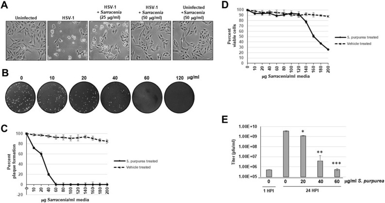
S. purpurea inhibited HSV-1 induced cytopathic effect and replication. (A) Vero cells were mock infected or infected with HSV-1 at a MOI = 5 and treated with 25 and 50 µg/ml of S. purpurea extract. The cell monolayers were photographed at 24 h.p.i. (B) Vero cells were infected with 100 pfu HSV-1 and treated with 0, 10, 20, 40, 60, or 120 µg/ml S. purpurea extract for 3 days. After 3 days, the cell monolayers were stained with crystal violet. (C) The plaque assay in (B) was repeated in the presence of the S. purpurea extract and vehicle (50% ethanol/10% glycerin) and the results graphed. Error bars indicate the standard deviation from three separate trials. (D) Vero cells were treated with increasing concentrations of S. purpurea extract or vehicle and cell viability measured at 24 h post-treatment and the results graphed. Error bars indicate the standard deviation from three separate trials. (E) Vero cells were infected with HSV-1 at a MOI = 5 and treated with 0, 20, 40, or 60 µg/ml of S. purpurea extract. Virus were harvested at 1 (for input virus titer) and 24 h.p.i. and titered. Statistical analysis was performed using a paired t-test. Samples with statistically significant deviation relative to the 0 μg/ml S. purpurea treatment are indicated with asterisks (*p < 0.05, **p < 0.01, ***p < 0.005).

