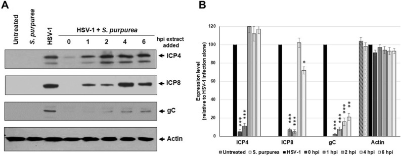
Figure 4.
S. purpurea reduced HSV-1 ICP4, ICP8, and gC protein levels in a time dependent manner. Vero cells were mock infected or infected with HSV-1 at a MOI = 5 in the presence or absence of S. purpurea (40 µg/ml) added at 0, 1, 2, 4, and 6 h.p.i. Cells were harvested at 16 h.p.i., lysed, separated by SDS-PAGE analyzed by Western blot with antibodies to HSV-1 ICP4, ICP8, gC and cellular actin. Actin was included as a standard loading control. (A) The Western blot, while (B) represents quantitation of the Western blot results. Error bars indicate the standard deviation from three separate analyses. Statistical analysis was performed using a paired t-test. Samples with statistically significant deviation relative to the untreated HSV-1 sample are indicated with asterisks (*p < 0.05, **p < 0.01, ***p < 0.005). Images of the full-length Western blots are included in the “Supplemental files”.

