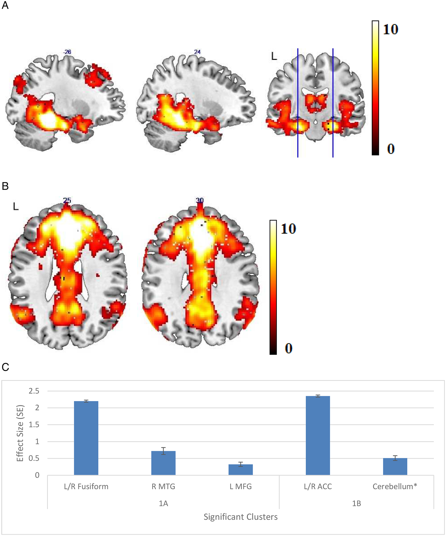
Figure 1.

(A) Areas of significant connectivity of the memory network for all participants in a whole brain correlation analysis with the left parahippocampal cortex (LPHC) as a seed region. (B) Areas of significant connectivity of the default mode network (DMN) for all participants in a whole brain correlation analysis with the medial prefrontal cortex (mPFC) as a seed region. (C) Effect size (average β value) by clusters with significant connectivity with seed regions with standard error (SE) plotted. Cluster regions are detailed in Table 2, labeled by the brain region containing peak voxel for the cluster.
* L: left, R: right, MTG: middle temporal gyrus, MFG: middle frontal gyrus, ACC: anterior cingulate cortex
