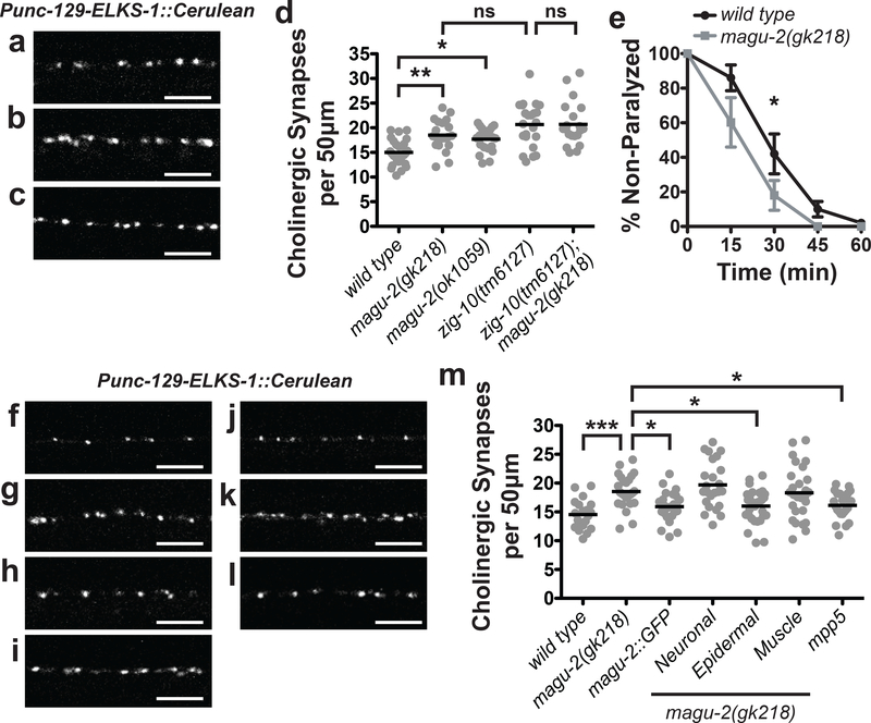
Figure 2. MAGU-2 functions in the epidermis to regulate cholinergic synapse density.
Representative images of cholinergic synapses labeled by ELKS-1::Cerulean in wild type (a), magu-2(gk218) (b), and magu-2(ok1059) (c) animals. Scale bars are 5 μm. (d) Quantification of synapses from the indicated genotypes of animals expressing ELKS-1::Cerulean in cholinergic neurons. Gray dots represent individual animals; the black lines indicate the means. *p<0.05; **p<0.01; ns= not significant. (e) Quantification of paralysis over time in the presence of 1 mM levamisole; *p<0.05. Representative images of cholinergic synapses labeled by ELKS-1::Cerulean in wild type (f), magu-2(gk218) (g), and magu-2(gk218); MAGU-2::GFP (h), magu-2(gk218); Neuronal::MAGU-2 (i), magu-2(gk218); Epidermal::MAGU-2 (j), magu-2(gk218); Muscle::MAGU-2 (k), and magu-2(gk218); Epidermal::mpp5 (l) animals. Scale bars are 5 μm. (m) Quantification of cholinergic synapses labeled by ELKS-1::Cerulean in wild type or magu-2(0) animals. Transgenic expression of MAGU-2 was achieved using its endogenous promoter, rgef-1 promoter for neurons, col-10 promoter for epidermis, or myo-3 promoter for muscle. Mouse mpp5 cDNA was expressed using the col-10 promoter. Gray dots represent individual animals; the black lines indicate the means; *p<0.05; ***p<0.001.

