
Figure 5.
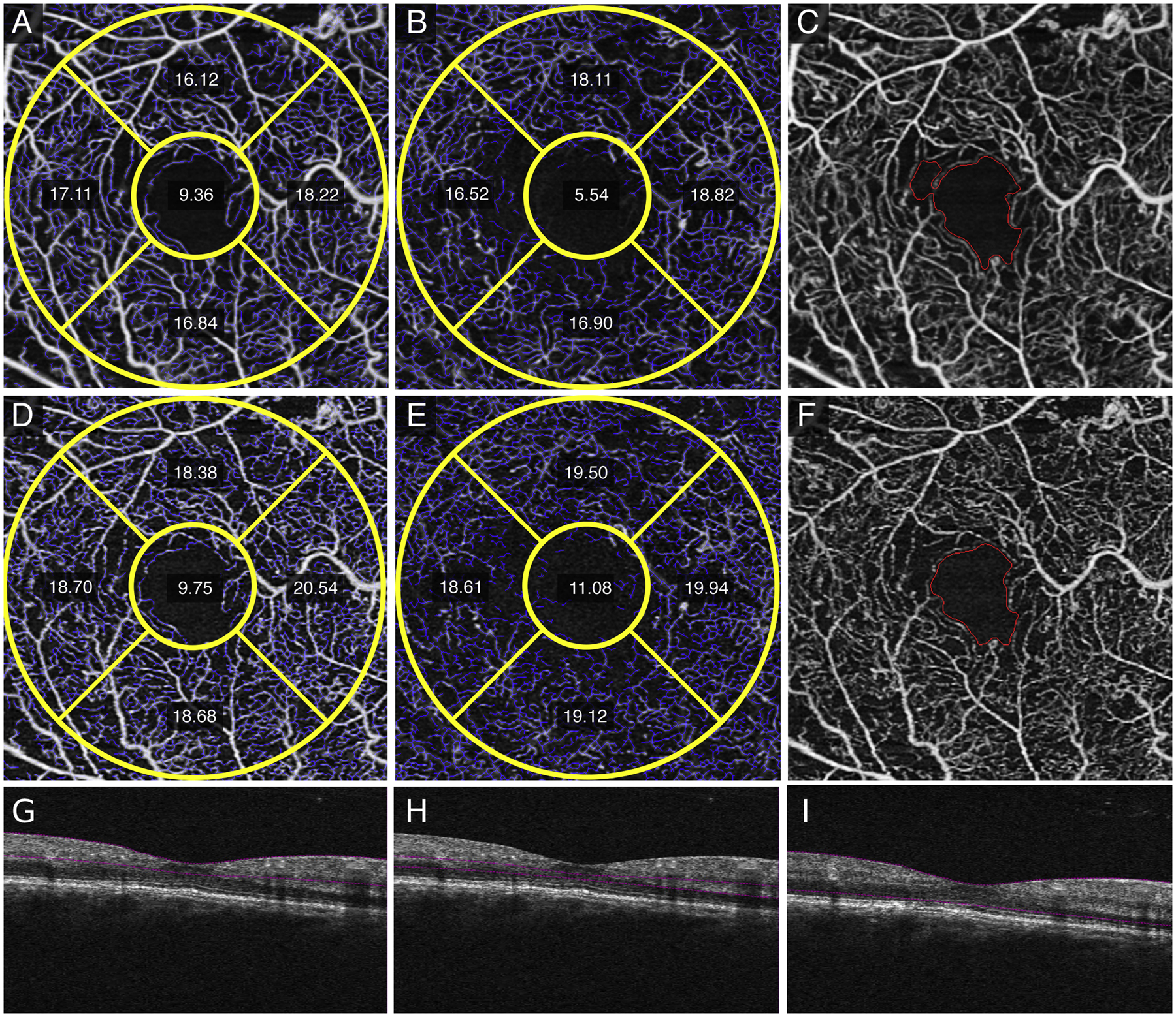
In a right eye with severe non-proliferative diabetic retinopathy, averaged superficial retinal layer (SRL) (A), deep retinal layer (DRL) (B), and fundus images with foveal avascular zone (FAZ) delineated (C) versus single image SRL (D), DRL (E), and FAZ (F) images show decreased vessel length density (VLD) measurements, improved vessel structure, and decreased noise, respectively. The structural non-averaged B-scans show the segmentation for the SRL (G), DRL (H), and full retina (I).