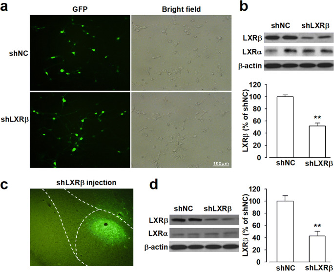
Fig. 4.
Knockdown of LXRβ expression by shRNA in cultured neurons and amygdala. (a) Cultured neurons were infected with LXRβ-shRNA (shLXRβ) or negative shRNA (shNC) for 72 h, and the efficiency of infection was observed as green fluorescence (GFP) by fluorescence microscope after infection with shRNA lentivirus (× 20), and corresponding bright field showed all the cultured neurons after infection. (b) Representative Western blots of LXRβ expression level of neurons that infected with shLXRβ or shNC for 72 h, β-actin served as internal control. shLXRβ decreased the expression level of LXRβ in cultured neurons. (c) Green fluorescence represented cells in the amygdala that were infected by shRNA lentivirus for 14 days (× 10). (d) Representative Western blots of LXRβ expression level in amygdala infected with shLXRβ or shNC for 14 days, β-actin served as internal control. shLXRβ decreased the expression level of LXRβ in the amygdala. Error bars represented SEM. n = 6, **p < 0.01, versus shNC-injected group

