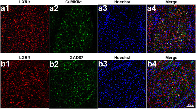
Fig. 8.
The cellular pattern of LXRβ colocalization in mice amygdala. The brain slices containing the amygdala were stained for (a) CAMKIIα+LXRβ. (b) GAD67 + LXRβ. LXRβ colocalized mainly with glutamatergic neurons (CAMKIIα positive), moderately with GABAergic neurons (GAD67 positive) in the amygdala. CAMKIIα and GAD67 showed in green, LXRβ showed in red, and Hoechst in blue. Scale bars = 100 μm

