Fig. 3.

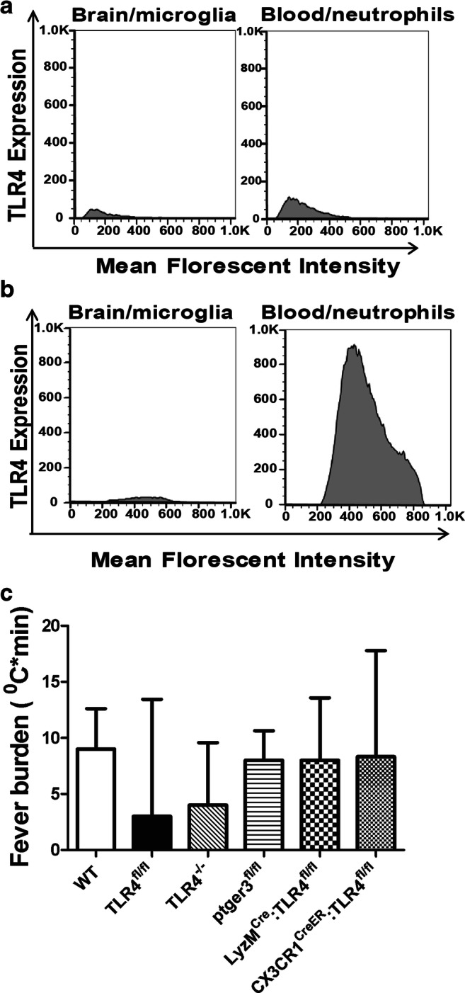
(A) Flow cytometry of brain homogenates and peripheral blood obtained from LyzMCre:TLR4fl/fl SAH mice was performed. Results are illustrated by the representative histogram. Blood injected into the brain was excluded from analysis by CD45.1+ selection, leaving only endogenous CD45.2+ cells. Microglia were sequentially gated: CD45.2+CD11bhiCX3CR1hiCD68intTmem119hi. This population was interrogated for TLR4 expression, which is represented by the brain/microglia histogram. Circulating monocytes and neutrophils were gated by size and granularity (forward and side scatter), and by CD11bhiCD68hi and Gr-1hi. As for the peripheral blood, TLR4 expression was subsequently interrogated and is represented by the blood/neutrophils histogram. Data demonstrated that TLR4 was not expressed in either microglia, or neutrophils (n = 3). (B) Similar flow and gating procedures were performed on the CX3Cr1CreER:TLR4fl/fl SAH mice. A representative histogram is shown. Data illustrated a lack of TLR4 expression in microglia, but normal expression of TLR4 in circulating neutrophils (n = 3 mice). (C) The fever burdens for wild-type C57BL/6 (WT), TLR4fl/fl, TLR4−/−, ptger3fl/fl, LyzMCre:TLR4fl/fl, and CX3Cr1CreER:TLR4fl/fl control mice injected with 60 μl of normal saline are shown with mean and standard error of the mean. All mice were injected with tamoxifen 1 week prior to the recording. Statistical significance was determined by one-way ANOVA and showed no differences between fever burdens of control NS injections between mice of different genotypes (n = 3 per group)
