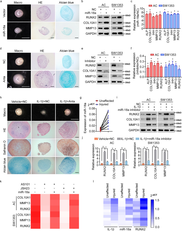
Fig. 2. IL-1β induced-miR-18a accelerates OA progression by promoting chondrocyte hypertrophy.
a Pellet cultures of MSCs expressing vector or miR-18a were induced to undergo chondrogenesis for 14 days and then induced for hypertrophic differentiation for another 14 days. Gross appearance, HE staining, and Alcian blue staining were evaluated. b, c WB b and qRT-PCR c to assess the expression of chondrocyte hypertrophy-related genes when miR-18a was ectopic expressed. d The effect of antagomir of miR-18a (Anta) on hypertrophy of chondrifying MSCs was assessed. e, f Expression of chondrocyte hypertrophy-related genes were determined by WB e and qRT-PCR f in AC and SW1353 cells transfected with inhibitor of miR-18a. g qRT-PCR shows miR-18a level in both injured cartilage and the corresponding paired unaffected cartilage. h The effect of miR-18a antagomir on IL-1β-enhanced hypertrophy of chondrifying MSCs was assessed. i, j Protein i and mRNA j levels of hypertrophy-related genes were analyzed in chondrocytes in response to IL-1β with or without inhibition of miR-18a. k qRT-PCR shows the effect of miR-18a ectopic expression on chondrocyte hypertrophy in cells treated with either AS101 or JSH23. l Expression of IL-1β, miR-18a, and RUNX2 were analyzed in injured cartilage and corresponding unaffected cartilage from OA patients. Scale bar in images of gross appearance in a, d, and h: 1 mm; other scale bars: 200 μm. Data in c, f, and j are presented as mean ± SD. *P < 0.05.

