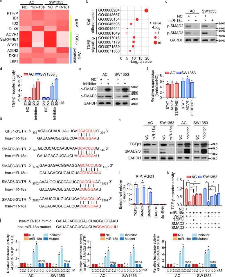
Fig. 3. miR-18a suppresses TGF-β signaling by targeting TGFβ1, SMAD2, and SMAD3.
a Expression of PTHrP and downstream genes of BMP, TGF-β, and Wnt/β-catenin signaling pathways was analyzed in miR-18a-overexpressed cells. b mRNA array was conducted in human ACs expressing control or miR-18a. And GO-enrichment analysis shows the correlation between expression of miR-18a and cell differentiation-associated signature or TGF-β signaling. c WB shows phosphorylation of SMAD2 and SMAD3 when miR-18a was overexpressed. d Dual-luciferase assays reveal TGF-β signaling activities. e, f Effect of inhibiting miR-18a on activity of TGF-β signaling was analyzed by WB e and qRT-PCR f. g Targetscan tool showing schematic representation of putative binding sites for miR-18a in 3′-UTRs of TGFβ1, SMAD2, and SMAD3. h WB analysis of the protein levels of TGFβ1 and SMAD2/3 in the indicated cells. i By immunoprecipitation against Ago1, RIP analysis reveals the interaction of miR-18a with the 3′-UTRs of TGFβ1, SMAD2, or SMAD3 mRNA to form miRNP complexes. IgG immunoprecipitation, as well as the interaction of miR-18a with GAPDH and 5s rRNA, were used as negative controls. j Luciferase assay of pGL3-TGFβ1-3′-UTR, pGL3-SMAD2-3′-UTR, and pGL3-SMAD3-3′-UTR reporters in the indicated cells, co-transfected with increasing amounts (20 and 50 nM) of the indicated oligonucleotides. The sequence of the miR-18a mutant is shown. k Effects of restored expression of TGFβ1, SMAD2, or SMAD3 in miR-18a-overexpressing cells on luciferase activities of the TGF-β reporter. Data in d, f, i, j, and k are presented as mean ± SD. *P < 0.05.

